SYSTEM SPECIFICATIONS Imaging System TriTomTM Repetition Rate 20 Hz PA Excitation Range 532 nm & 650-1300 (2300) nm
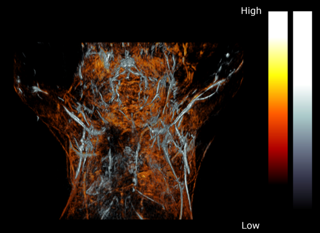
Figure 1: PAT composite image of the upper torso and brain of a female BALB/c mouse acquired post-mortem with 532 nm excitation (orange) and 750 nm (grey).
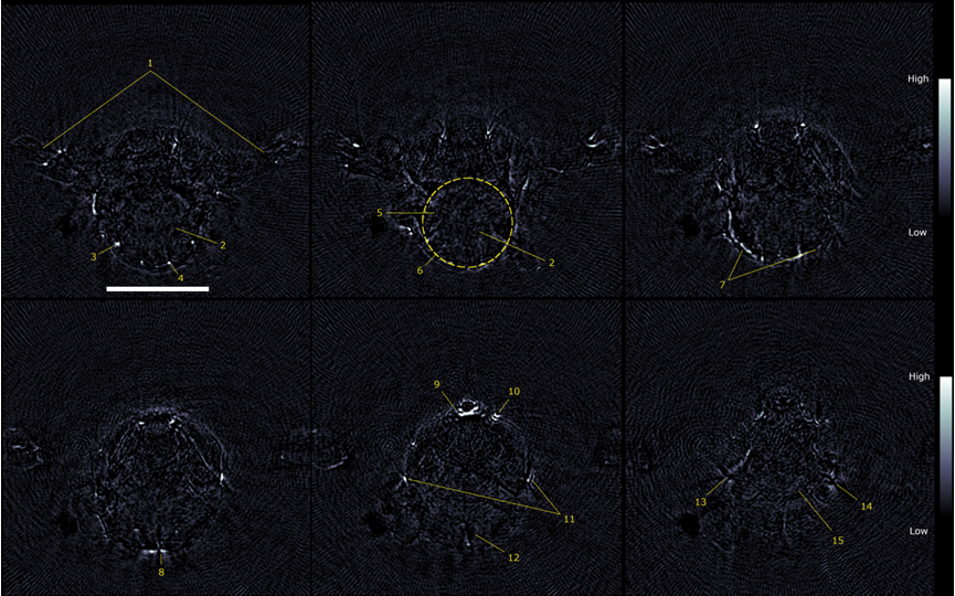
Figure 2: 2D transverse slices of the 750 nm PAT scans acquired at several vertical displacements. (1) arms, (2) cerebellum, (3) auricular artery, (4) cerebral artery, (5) medulla, (6) outline of brain, (7) transverse sinus, (8) confluence of sinus, (9) sublingual vein, (10) facial vein, (11) superficial temporal vein, (12) subarachnoid space, (13) right eye, (14) left eye, (15) optic track. Scale bar = 5 mm.
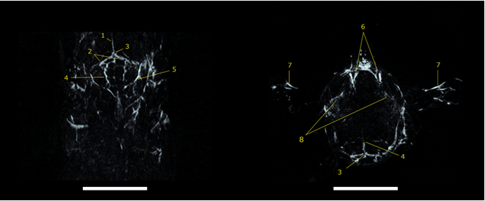
Figure 3: 10 mm thick coronal (left) and transverse (right) maximum intensity projection slabs of a PAT volume reconstructed from the 750 nm scan. (1) Superior sagittal sinus, (2) transverse sinus, (3) confluence of sinus, (4) cerebral artery, (5) auricular artery, (6) jugular vein, (7) brachial artery, (8) ophthalmic artery. Scale bar = 5 mm.
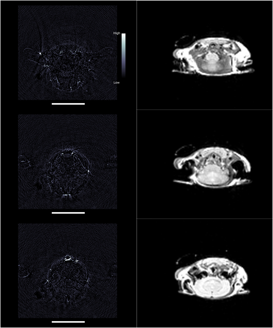
Figure 4: PAT (left) and MRI (right) scans of two female BALB/c mice demonstrating the anatomical and functional information acquired with complementary imaging modalities. Scale bars = 10 mm.